
Most discussions of mast cell biology begin and end with histamine. The traditional model is familiar: an allergen activates IgE antibodies, IgE binds to the FcεRI receptor on mast cells, histamine is released, and allergic symptoms follow. This framework has helped us understand allergies, asthma, and anaphylaxis for decades.
The problem is that mast cells are far more complex. Modern immunology has revealed that mast cells express dozens of receptors capable of responding to signals from the immune system, nervous system, endocrine system, microbiome, and surrounding tissue environment. The diagram below, illustrates the remarkable complexity of mast cell signaling and the vast network of inputs capable of activating these cells.
Rather than acting as simple histamine factories, mast cells function as highly sophisticated environmental sensors embedded within the connective tissues of the body.
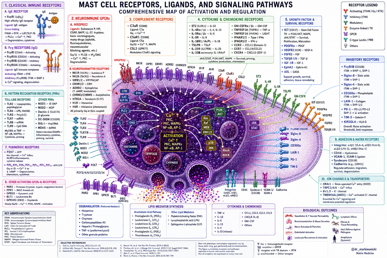
Figure concept developed by Dr. Mark Iwanicki and rendered with assistance from ChatGPT (OpenAI).
The Major Mast Cell Pathways We Already Know
The best understood mast cell pathway remains the IgE receptor FcεRI. This pathway drives classical allergic reactions and has been the focus of much of modern allergy medicine.
Other clinically important pathways include:
Histamine receptors (targeted by antihistamines)
Leukotriene pathways (targeted by medications such as montelukast)
KIT signaling pathways that regulate mast cell growth and survival
IgE itself, which can be targeted with biologic therapies such as omalizumab
These interventions can be extraordinarily valuable and have transformed the management of many mast cell disorders. Yet even these pathways represent only a fraction of what mast cells actually do.
The Neuroimmune Mast Cell
Mast cells sit in close proximity to autonomic nerve endings throughout the body.
Through receptors such as MRGPRX2, mast cells can respond directly to neuropeptides including Substance P, CGRP, and other signals released by sensory nerves. This creates a two-way conversation between the nervous system and immune system.
Stress, trauma, chronic pain, neuroinflammation, and autonomic dysfunction can all influence mast cell activity through these pathways. This helps explain why many patients experience mast cell symptoms that cannot be fully explained by allergies alone.
The Gut-Mast Cell Axis
Mast cells are highly concentrated within the intestinal lamina propria, immediately beneath the epithelial barrier.
This strategic location allows them to monitor the interface between the outside world and the internal environment. Bacterial products such as lipopolysaccharide (LPS) can activate mast cells through Toll-like receptors, particularly TLR4. Dysbiosis, increased intestinal permeability, chronic infection, and microbial imbalance may therefore contribute to ongoing mast cell activation even in the absence of traditional allergens.
In many ways, mast cells serve as guardians of the gut barrier.
Hormones and Mast Cells
Mast cells are also responsive to hormonal signals. Estrogen receptors have been identified on mast cells, and estradiol has been shown to influence mast cell activation and mediator release. This may help explain why some individuals experience symptom fluctuations throughout the menstrual cycle, during pregnancy, or during perimenopause. For many patients, hormones are not separate from mast cell biology. They are part of it.
The Complement Connection
The complement system provides another powerful mechanism of mast cell activation.
Complement fragments such as C3a and C5a are capable of directly activating mast cells and amplifying inflammatory responses. These pathways may become particularly relevant in chronic infections, mold-related illness, tissue injury, and autoimmune conditions. Increasingly, mast cells appear less like isolated immune cells and more like central communication hubs integrating signals from multiple biological systems simultaneously.
The Matrix Environment & The Mast Cell
What is often overlooked is where mast cells actually live.
Mast cells do not float freely through the body. They reside within the extracellular matrix, the fluid-filled connective tissue network that surrounds every cell.
This matrix contains collagen fibers, ground substance, capillaries, lymphatic vessels, fibroblasts, autonomic nerve endings, immune cells, nutrients, oxygen, signaling molecules, and metabolic waste products. It is the environment through which every cellular conversation occurs.
From this perspective, mast cells are not simply responding to allergens.
They are responding to the condition of the terrain around them. Research has demonstrated that mast cells can be activated by:
Hypoxia
Oxidative stress
Acidic tissue environments
Extracellular ATP released from damaged cells
Mechanical stress
Fibrotic remodeling
Inflammatory cytokines
Microbial products
Tissue injury
Viewed collectively, these signals paint a fascinating picture. The mast cell appears designed to monitor the health of the local tissue environment and respond whenever that environment becomes threatened.
A Matrix Medicine Perspective
From a Matrix Medicine perspective, many of these processes can be understood through the lens of matrix physiology. When microcirculation becomes impaired, oxygen delivery falls. When lymphatic drainage slows, inflammatory mediators accumulate. When tissues become chronically inflamed, extracellular pH shifts and oxidative stress rises. When microbial products escape the gut barrier, mast cells are among the first cells to encounter them.
Rather than viewing these events as isolated phenomena, Matrix Medicine sees them as manifestations of a disturbed extracellular environment.
This concept is sometimes described as matrix congestion.
Not congestion in the simplistic sense of fluid retention, but a broader disturbance in extracellular physiology characterized by impaired oxygen diffusion, altered fluid dynamics, accumulation of inflammatory signaling molecules, oxidative stress, altered pH regulation, and disruption of normal cell-to-cell communication.
Under these conditions, mast cells may be doing exactly what they evolved to do: respond to danger.
Why This Changes Treatment
Most conventional therapies focus on blocking mast cell mediators after they are released or preventing mast cells from releasing them in the first place. These approaches can be extremely helpful and often necessary. However, they do not directly address the tissue environment generating many of the signals that activate mast cells.
This is where Matrix Medicine takes a different approach.
Rather than focusing exclusively on the mast cell itself, treatment seeks to improve the environment in which the mast cell resides. This includes supporting lymphatic flow and downstream primary detoxification organs (Liver, Kidney, Intestines), alkalinizing the terrain, improving microcirculation, restoring gut barrier integrity, reducing inflammatory burden, improving oxygen delivery, regulating autonomic function, all techniques aimed at optimizing extracellular matrix physiology.
Within European Biological Medicine, drainage therapies have long been used to support these goals. Practitioners frequently observe that remedies from companies such as GUNA, Pekana, Heel, and others can produce profound clinical improvements in patients with chronic inflammatory and mast cell-related presentations.
These therapies are not generally designed to target mast cells directly. Instead, they are intended to support the extracellular matrix, lymphatic system, organs of elimination, and broader terrain in which immune regulation occurs.
Whether viewed through the language of drainage, matrix physiology, tissue microenvironments, or systems biology, the underlying principle is similar: changing the environment often changes the behavior of the cells living within it.
Looking Beyond Histamine
The future of mast cell medicine may not lie solely in discovering new receptor blockers.
It may also lie in understanding why those receptors are being activated in the first place.
Mast cells are not merely allergy cells. They are environmental sensors embedded within the extracellular matrix, continuously interpreting signals from the nervous system, immune system, microbiome, endocrine system, and the terrain that surrounds them. Viewed through this lens, the mast cell becomes less an enemy to suppress and more a messenger reporting on the state of the environment in which it lives.
This perspective does not replace conventional approaches to mast cell disorders. Histamine blockers, leukotriene inhibitors, mast cell stabilizers, biologics, and other targeted therapies can be invaluable tools. But they represent only one side of the equation.
The other side is the environment in which the mast cell resides. As our understanding of mast cell biology continues to evolve, the extracellular matrix may prove to be one of the most important and overlooked pieces of the puzzle. The mast cell is not simply responding to allergens. It is responding to its surroundings. Understanding those surroundings may ultimately help us understand why chronic mast cell activation develops in the first place.
Want to Learn More?
For Practitioners
If this article resonates with you and you would like to learn how to apply Matrix Medicine principles in clinical practice, including extracellular matrix physiology, drainage, terrain regulation, and the role of the matrix in chronic inflammatory conditions, I invite you to watch my free masterclass:
The Matrix Reset Masterclass
https://courses.drmarkiwanicki.com/the-matrix-reset-masterclass
In this training, I explore the extracellular matrix as the missing physiological layer beneath many chronic conditions and explain how Matrix Medicine can be incorporated into everyday clinical practice.
For Patients
If you are dealing with chronic inflammatory symptoms, mast cell activation, gut dysfunction, environmental sensitivities, complex chronic illness, or other unresolved health concerns and would like personalized support, you can schedule an online consultation here:
https://drmarkiwanicki.com/work-with-me
References
Krystel-Whittemore M, Dileepan KN, Wood JG. Mast Cell: A Multi-Functional Master Cell. Front Immunol. 2016.
Roy S, Na Ayudhya CC, Thapaliya M, Ali H. Multifaceted MRGPRX2: New Insight into the Role of Mast Cells in Health and Disease. J Allergy Clin Immunol. 2021.
Nagata Y, Suzuki R. FcεRI: A Master Regulator of Mast Cell Functions. Cells. 2022.
Espinosa-Riquer ZP, Segura-Villalobos D, et al. Signal Transduction Pathways Activated by Innate Immunity in Mast Cells. Cells. 2020.
Ali H. MRGPRX2 and IgE-Independent Mast Cell Activation. Front Immunol. 2021.
Marshall JS, Portales-Cervantes L, Leong E. Mast Cell Responses to Viruses and Pathogen Products. Int J Mol Sci. 2019.
Pundir P, Liu R, Vasavda C, et al. A Connective Tissue Mast Cell-Specific Receptor Detects Bacterial Quorum-Sensing Molecules and Mediates Antibacterial Immunity. Cell Host Microbe. 2019.
Pastwińska J, Agier J, Dastych J, Brzezińska-Błaszczyk E. Hypoxia Regulates Human Mast Cell Adhesion to Fibronectin. Mediators Inflamm. 2020.
Möllerherm H, von Köckritz-Blickwede M, Branitzki-Heinemann K. Hypoxia Modulates the Response of Mast Cells to Bacterial Stimuli. Front Immunol. 2017.
Gulliksson M, Carvalho RF, Ulleras E, Nilsson G. Mast Cell Survival and Mediator Secretion in Response to Hypoxia. PLoS One. 2010.
Wang X, Li X, Ya F, et al. Hypoxic Mast Cells Accelerate Fibrosis and Tissue Remodeling. Int J Mol Med. 2020.
Hügle T. Beyond Allergy: The Role of Mast Cells in Fibrosis. Swiss Med Wkly. 2014.
Valent P, Akin C, Hartmann K, et al. Mast Cells as a Unique Hematopoietic Lineage and Cell System. Theranostics. 2020.
Sandhu JK, Kulka M. Decoding Mast Cell-Microglia Communication in Neurodegenerative Disease. Int J Mol Sci. 2021.
Komi DEA, Kuebler WM. Significance of Mast Cell Extracellular Traps in Microbial Defense. Clin Rev Allergy Immunol. 2022.
Thapaliya M, Roy S, Ali H. Regulation of FcεRI and MRGPRX2-Mediated Responses in Mast Cells. Front Immunol. 2023.
Theoharides TC. Mast Cells and Stress: Neuroimmune Interactions in Health and Disease. J Clin Psychopharmacol.
Theoharides TC, Stewart JM, Taracanova A, et al. Neuroendocrinology of Mast Cells. Mol Neurobiol.
Vliagoftis H, Dimitriadou V, Boucher W, et al. Estradiol Augments Mast Cell Activation and Mediator Release. Endocrinology.
Galli SJ, Tsai M. Mast Cells in Health and Disease. N Engl J Med.